40 microscope images with labels
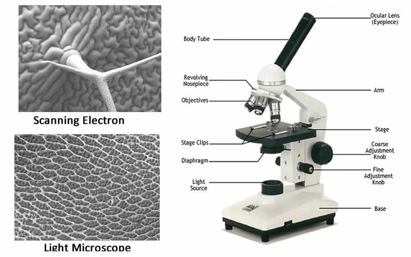
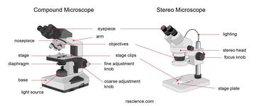
Light Microscope- Definition, Principle, Types, Parts, Labeled Diagram ... The image formed is a fluorochrome-labeled image from the emitted light The principle behind this working mechanism is that the fluorescent microscope will expose the specimen to ultra or violet or blue light, which forms an image of the specimen that is emanated by the fluorescent light. How to Use the Microscope - The Biology Corner Stereoscope - this microscope allows for binocular (two eyes) viewing of larger specimens. Scanning Electron Microscope - allow scientists to view a universe too small to be seen with a light microscope. SEMs do not use light waves; they use electrons (negatively charged electrical particles) to magnify objects up to two million times.
How Magnets Work | HowStuffWorks Apr 02, 2007 · Iron filings beautifully show off the opposing fields of the same poles of two bar magnets. Spencer Grant/Photographer's Choice RF/Getty Images. You probably know that magnets attract specific metals and they have north and south poles. Opposite poles attract each other while like poles repel each other. Magnetic and electrical fields are related, and magnetism, along with gravity and strong ...

Microscope images with labels
Skin Images Labeled | Virtual Anatomy Lab VAL - ncccval Body cavities, planes, and regions. Body Images Labeled. Body Images Unlabeled. Histology. Epithelium Images Labeled. Epithelium Images Unlabeled. Connective Tissue Images Labeled. Connective Tissue Images Unlabeled. Microscope. Microscope Labeled Pictures, Images and Stock Photos Browse 49 microscope labeled stock photos and images available, or start a new search to explore more stock photos and images. Newest results Fluorescent Imaging immunofluorescence of cancer cells growing... Microscope diagram vector illustration. Labeled zoom instrument... Microscope diagram vector illustration. Label the microscope — Science Learning Hub All microscopes share features in common. In this interactive, you can label the different parts of a microscope. Use this with the Microscope parts activity to help students identify and label the main parts of a microscope and then describe their functions. Drag and drop the text labels onto the microscope diagram.
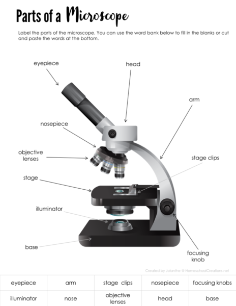
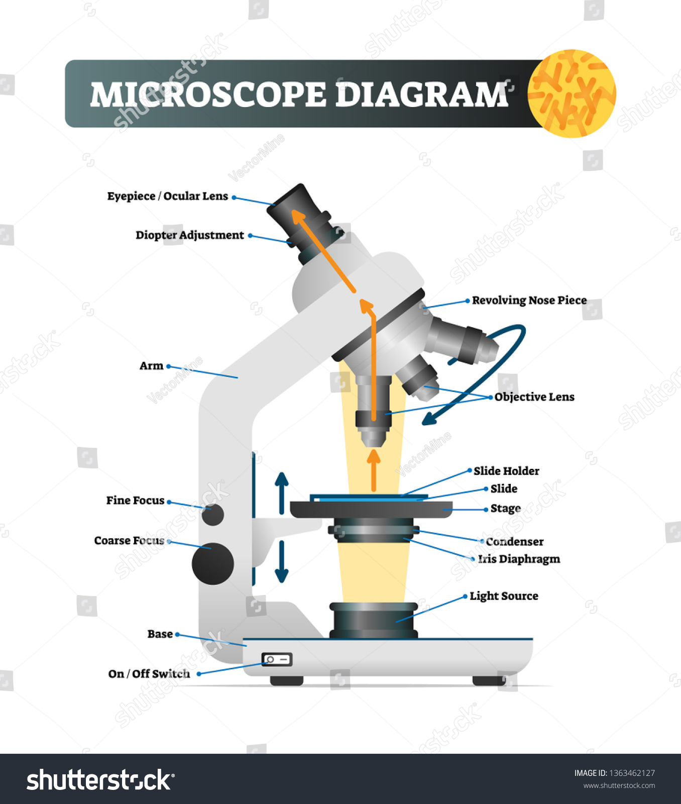
Microscope images with labels. 400+ Free Microscope & Bacteria Images - Pixabay 400+ Free Microscope & Bacteria Images - Pixabay / 5 ‹ › SafeSearch Most Relevant Images Orientation Size Color Published date 413 Free images of Microscope Related Images: bacteria laboratory science research scientist biology lab chemistry microbiology Find your perfect microscope image. Free pictures to download and use in your next project. Amazon.com: 58-pcs Microscope Kit for Kids 5-7 8-12, 100X ... Aug 28, 2021 · VALUE FOR YOU MONEY - Comes with 58-pcs microscope kit: 1 x Microscope, 12 x Random Specimens, 1 x Specific Specimen, 31 x Blank Plastic Slides, 1 x Tweezer, 1 x Mixing Plate, 1 x Dropper, 1 x Scalpel, 1 x Stirring Rod, 4 x Mini Volumetric Bottles, 2 x Blank Labels Boxes, 1 x Mobile Phone Holder, 1 x ABS Carry Box, 1 x Instruction. Parts of a microscope with functions and labeled diagram - Microbe Notes Parts of a microscope with functions and labeled diagram September 17, 2022 by Faith Mokobi Having been constructed in the 16th Century, Microscopes have revolutionalized science with their ability to magnify small objects such as microbial cells, producing images with definitive structures that are identifiable and characterizable. Labeling the Parts of the Microscope | Microscope World Resources Labeling the Parts of the Microscope This activity has been designed for use in homes and schools. Each microscope layout (both blank and the version with answers) are available as PDF downloads. You can view a more in-depth review of each part of the microscope here. Download the Label the Parts of the Microscope PDF printable version here.
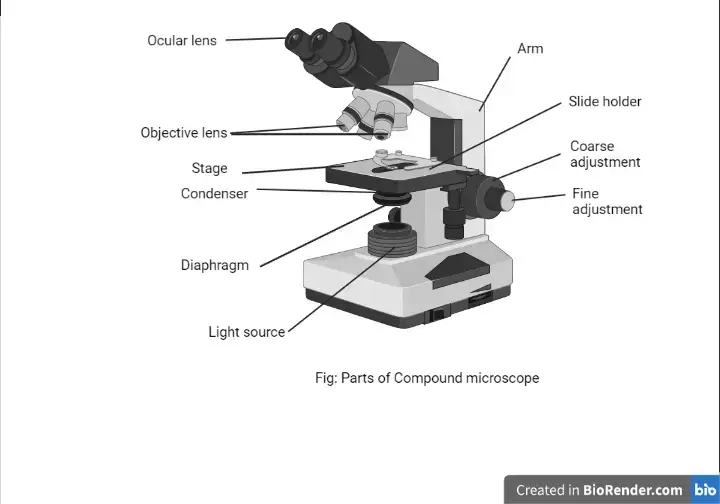
Mitosis Images Labeled | Virtual Anatomy Lab VAL - ncccval Endocrine Rabbit Dissection Unlabeled. Cardiovascular. Cardiovascular Histology Labeled. Cardiovascular Histology Unlabeled. Cardiovascular Models Labeled. Cardiovascular Models Unlabeled. Cardiovascular Sheep Heart Dissect-L. Cardiovascular Sheep Heart Disect-U. Cardiovascular Cat Dissection Labeled. Compound Microscope Parts, Functions, and Labeled Diagram Compound Microscope Definitions for Labels. Eyepiece (ocular lens) with or without Pointer: The part that is looked through at the top of the compound microscope. Eyepieces typically have a magnification between 5x & 30x. Monocular or Binocular Head: Structural support that holds & connects the eyepieces to the objective lenses. Explanation and Labelled Images - New York Microscope Company The samples are labeled with fluorophore where they absorb the high-intensity light from the source and emit a lower energy light of longer wavelength. The resulting fluorescent light is then separated from the surrounding radiation with filters, allowing the observer to see only the fluorescing material. Parts of the Microscope with Labeling (also Free Printouts) Microscopes are specially created to magnify the image of the subject being studied. This exercise is created to be used in homes and schools. the microscope layout, including the blank and answered versions are available as pdf downloads. Click to Download : Label the Parts of the Microscope (A4) PDF print version.
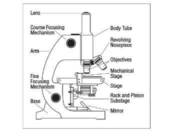
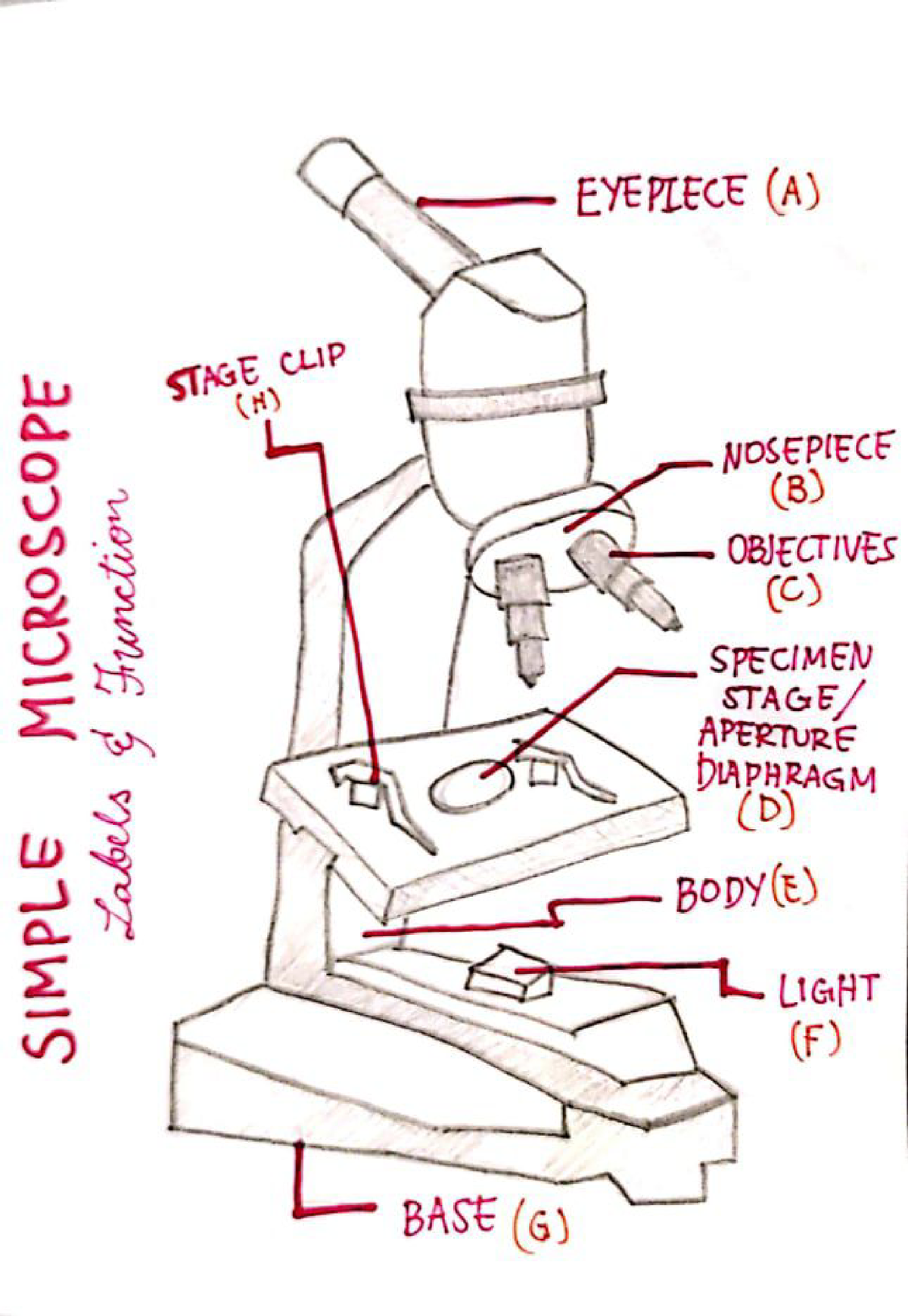
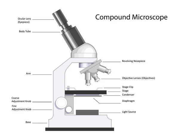
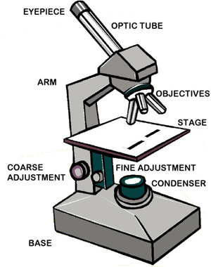
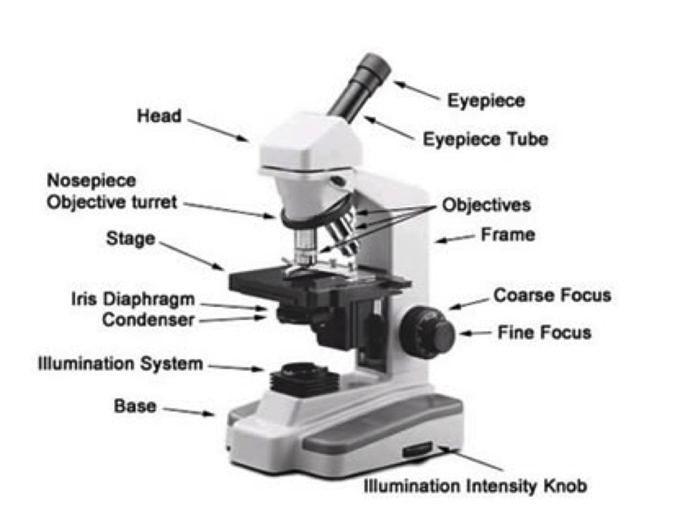
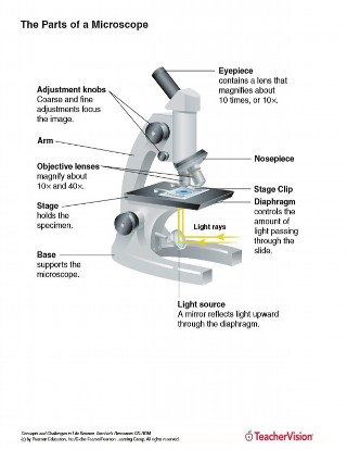
National Geographic Dual LED Student Microscope Aug 07, 2017 · Buy NATIONAL GEOGRAPHIC Dual LED Student Microscope - 50+ pc Science Kit with 10 Prepared Biological & 10 Blank Slides, Lab Shrimp Experiment, Perfect for School Laboratory, Homeschool & Home Education: Microscopes - Amazon.com FREE DELIVERY possible on eligible purchases Stock Quotes, Business News and Data from Stock Markets | MSN ... Get the latest headlines on Wall Street and international economies, money news, personal finance, the stock market indexes including Dow Jones, NASDAQ, and more. Be informed and get ahead with ... Microscope Parts and Functions Body tube (Head): The body tube connects the eyepiece to the objective lenses. Arm: The arm connects the body tube to the base of the microscope. Coarse adjustment: Brings the specimen into general focus. Fine adjustment: Fine tunes the focus and increases the detail of the specimen. Nosepiece: A rotating turret that houses the objective lenses. Microscope Types (with labeled diagrams) and Functions The working principle of a simple microscope is that when a lens is held close to the eye, a virtual, magnified and erect image of a specimen is formed at the least possible distance from which a human eye can discern objects clearly. Simple microscope labeled diagram Simple microscope functions It is used in industrial applications like:
Learning Task 3: Label Me!Label The Parts of the Microscope ...
Microscope Stock Photos, Pictures & Royalty-Free Images - iStock Browse 201,051 microscope stock photos and images available, or search for magnifying glass or microscope isolated to find more great stock photos and pictures. Microscope icon. Medical diagnostics, laboratory, research, education, study. Vector illustration. Equipment for medical research, laboratories.
Parts of a Microscope - Free Printable
Microscope Labeling - The Biology Corner The google slides shown below have the same microscope image with the labels for students to copy. I often spend the first day walking students through the steps and having them look at a single slide as we do the steps. Students are often very enthusiastic about using microscopes and will try to start with the high power objective.

Educational / Hobby Microscope (BE211A Eco-Bino-LED)
Connective Tissue Images Labeled | Virtual Anatomy Lab VAL - ncccval Body Images Labeled. Body Images Unlabeled. Histology. Epithelium Images Labeled. Epithelium Images Unlabeled. Connective Tissue Images Labeled. Connective Tissue Images Unlabeled. Microscope. Microscope Images Labeled.

Labeling the Parts of the Microscope | Microscope activity ...
Microscope Images Unlabeled | Virtual Anatomy Lab VAL - ncccval Body cavities, planes, and regions. Body Images Labeled. Body Images Unlabeled. Histology. Epithelium Images Labeled. Epithelium Images Unlabeled. Connective Tissue Images Labeled. Connective Tissue Images Unlabeled. Microscope.
Parts of a Microscope with Their Functions – Microbe Online
Questia - Gale Questia. After more than twenty years, Questia is discontinuing operations as of Monday, December 21, 2020.

Compound Microscope Parts – Labeled Diagram and their ...
Laser Scanning Confocal Microscopy | Nikon’s MicroscopyU This tutorial explores imaging of specimens with a Nikon PCM 2000 laser scanning confocal microscope by creating virtual control systems that simulate how the actual microscope operates. All specimens contained in this tutorial were imaged with the Nikon instrument and are presented as successive z-axis optical sections obtained from the ...
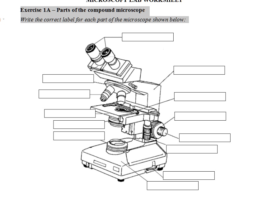
SOLVED: Exercise 1A Parts ofthe compound microscope Write the ...
Compound Microscope Parts - Labeled Diagram and their Functions The eyepiece (or ocular lens) is the lens part at the top of a microscope that the viewer looks through. The standard eyepiece has a magnification of 10x. You may exchange with an optional eyepiece ranging from 5x - 30x. [In this figure] The structure inside an eyepiece. The current design of the eyepiece is no longer a single convex lens.
microscope | The Biology Corner
Microscope Labeling Game - PurposeGames.com About this Quiz. This is an online quiz called Microscope Labeling Game. There is a printable worksheet available for download here so you can take the quiz with pen and paper. This quiz has tags. Click on the tags below to find other quizzes on the same subject. Science.
Microscope with labels picture
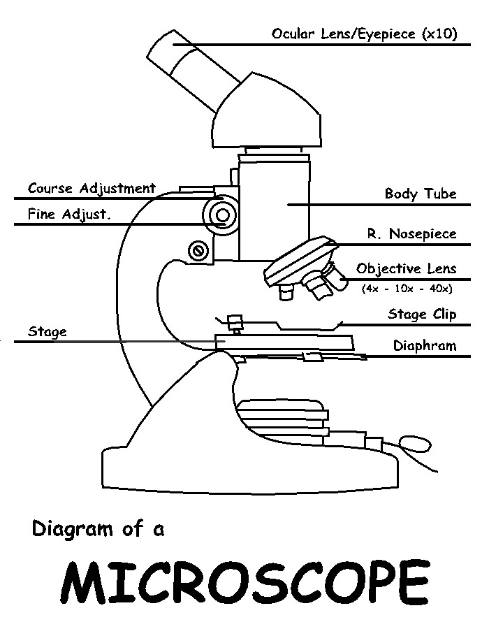
PDF Label parts of the Microscope Label parts of the Microscope: . Created Date: 20150715115425Z
Color the Microscope Parts
Label the microscope — Science Learning Hub All microscopes share features in common. In this interactive, you can label the different parts of a microscope. Use this with the Microscope parts activity to help students identify and label the main parts of a microscope and then describe their functions. Drag and drop the text labels onto the microscope diagram.

Microscope Labels Flashcards | Quizlet
Microscope Labeled Pictures, Images and Stock Photos Browse 49 microscope labeled stock photos and images available, or start a new search to explore more stock photos and images. Newest results Fluorescent Imaging immunofluorescence of cancer cells growing... Microscope diagram vector illustration. Labeled zoom instrument... Microscope diagram vector illustration.

Microscopes / Group 5
Skin Images Labeled | Virtual Anatomy Lab VAL - ncccval Body cavities, planes, and regions. Body Images Labeled. Body Images Unlabeled. Histology. Epithelium Images Labeled. Epithelium Images Unlabeled. Connective Tissue Images Labeled. Connective Tissue Images Unlabeled. Microscope.

Microscope With Labels free vector | Download it now!
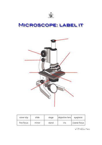
Microscope: label it | Teaching Resources
Solved] Can you identify the stage clip and the coaxial stage ...
Parts of Stereo Microscope (Dissecting microscope) – labeled ...
Microscope- Simple-AND Compound-WITH- Label - BS in Education ...
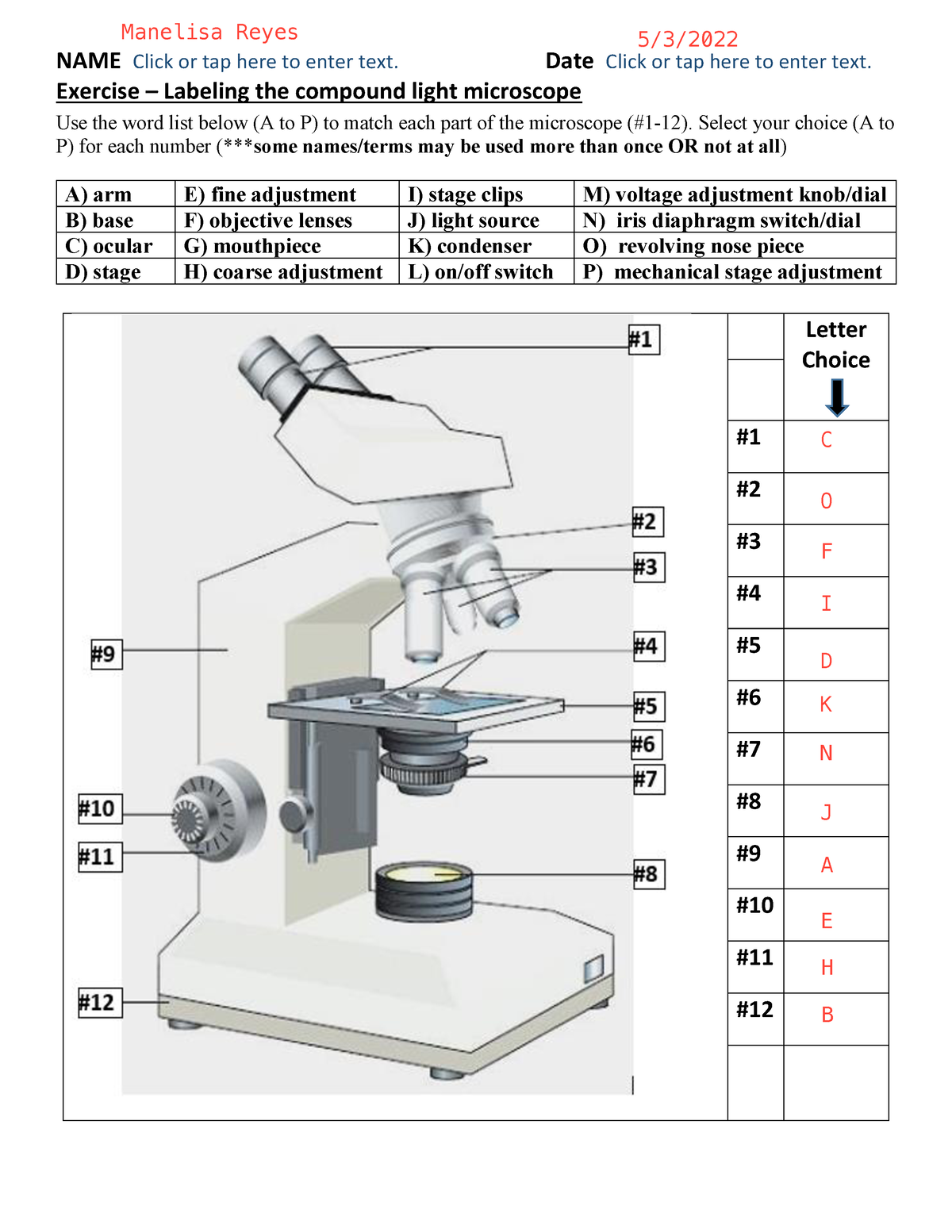
BIO 101 parts of the microscope to label - NAME Click or tap ...

This is a common compound microscope. Label its parts from A ...

Binocular Microscope Parts Quiz

Microscope labeling

Parts of a Microscope - SmartSchool Systems

Label Microscope Diagram - EnchantedLearning.com
Microscope slide Vector Art Stock Images | Depositphotos
Label microscope - Teaching resources

Label The Microscope Parts! Diagram | Quizlet

This is a common compound microscope. Label its parts from A ...

Labeling a Microscope Free Worksheet Pack
Comparing and Contrasting the Different Parts of the Microscope

microscope drawing with label - Clip Art Library

Week 1: Microscope Usage & Snowflake Preservation
Monday 10/19/15 AIM: how do the parts of the compound light ...

Meiji MT6500 Series PCM NIOSH 7400 Asbestos Microscope
Label microscope - Teaching resources
4,875 Microscope Labeled Images, Stock Photos & Vectors ...
The Parts of a Microscope (Labeled) Printable Printable (6th ...
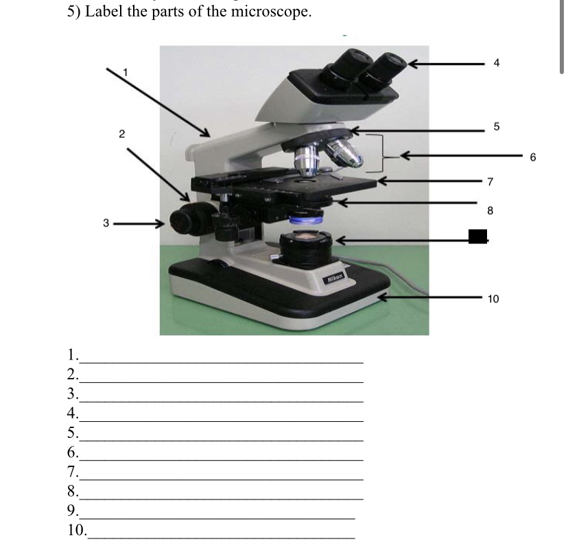
Answered: 5) Label the parts of the microscope. 1… | bartleby

Microscope « KaiserScience

What is a Compound Microscope? | Microscope World Blog
Free Microscope Drawing, Download Free Microscope Drawing png ...
Post a Comment for "40 microscope images with labels"