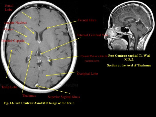
41 brain mri with labels
Labeled MRI Brain Scans - Neuromorphometrics We can also label scans that you provide and we are very interested in labeling white matter anatomy as seen in diffusion-weighted MRI scans. If you want an aggregate version of our data, we can provide it as a probabilistic atlas. The cost to label a single scan is $2449 (USD). High resolution automated labeling of the hippocampus and amygdala ... In this study, we used a recently developed 3D convolutional neural network (CNN) architecture "DeepMedic" (Kamnitsas et al., 2017), originally developed to label brain lesions, to train a model to label hippocampi and amygdala on whole brain 700 μm isotropic MP2RAGE MRI acquired at 7T in healthy controls and individuals with epilepsy ...
brain midsagittal view labels - Sagittal Section Of Brain Labeled | mri ... Brain Midsagittal View Labels images that posted in this website was uploaded by Media.nbcmontana.com. Brain Midsagittal View Labels equipped with a HD resolution 1064 x 851.You can save Brain...

Brain mri with labels
Atlas of BRAIN MRI - W-Radiology Brain magnetic resonance imaging (MRI) is a common medical imaging method that allows clinicians to examine the brain's anatomy (1). It uses a magnetic field and radio waves to produce detailed images of the brain and the brainstem to detect various conditions (2). CaseStacks.com - MRI Brain Anatomy Labelled radiographs and CT/MRI series teaching anatomy with a level of detail appropriate for medical students and junior residents. Pelvis Pelvic MRI anatomy Chest Chest radiograph & CT anatomy Body Abdominal CT anatomy Cardiac Cardiac CT anatomy Brain Brain & calvarial anatomy on CT/MRI Cranial Nerves Cranial nerves on MRI 101 Labeled Brain Images and a Consistent Human Cortical Labeling ... Labeled anatomical subdivisions of the brain enable one to quantify and report brain imaging data within brain regions, which is routinely done for functional, diffusion, and structural magnetic resonance images (f/d/MRI) and positron emission tomography data.
Brain mri with labels. brain and parts labeled brain and parts labeled brain and parts labeled Mri eye anatomy muscles scan sciencephoto. Diencephalon and brain stem: unit 4, group 3. Brain sagittal anatomy human section labeled cat robotspacebrain cut diagram parts physiology medical mid function mri structure labels drawing fig brain and parts labeled Researchers automate brain MRI image labeling, more than 100,000 exams ... Researchers have automated brain MRI image labeling, needed to teach machine learning image recognition models, by deriving important labels from radiology reports and accurately assigning them to... Brain lobes - annotated MRI | Radiology Case | Radiopaedia.org Debowski, M. Brain lobes - annotated MRI. Case study, Radiopaedia.org. (accessed on 17 Jul 2022) Automated MRI image labelling processes 100,000 brain exams in under 30 ... Researchers from the School of Biomedical Engineering & Imaging Sciences at King's College London have automated brain MRI image labeling, needed to teach machine learning image recognition models,...
Cross-sectional anatomy of the brain - e-Anatomy - IMAIOS We created a brain atlas that is an interactive tool for studying the conventional anatomy of the normal brain based on a magnetic resonance imaging exam of the axial brain. Anatomical structures and specific areas are visible as interactive labeled images. Cross sectional anatomy: MRI of the brain. An MRI was performed on a healthy subject ... brain anatomy | MRI coronal brain anatomy | free MRI cross sectional ... This MRI brain coronal cross sectional anatomy tool is absolutely free to use. Use the mouse scroll wheel to move the images up and down alternatively use the tiny arrows (>>) on both side of the image to move the images.>>) on both side of the image to move the images. MRI anatomy | free MRI axial brain anatomy MRI anatomy | free MRI axial brain anatomy This MRI brain cross sectional anatomy tool is absolutely free to use. Use the mouse scroll wheel to move the images up and down alternatively use the tiny arrows (>>) on both side of the image to move the images. MRI Brain Atlas This web app Atlas is intended for veterinary students and radiologists seeking quick access to canine brain anatomy through a mobile device. Via a toggle button, either MRI images or approximately comparable Brain Transection images may be viewed with or without labels. Navigation & Labels.
Brain: Atlas of human anatomy with MRI - e-Anatomy - IMAIOS MRI Atlas of the Brain. This page presents a comprehensive series of labeled axial, sagittal and coronal images from a normal human brain magnetic resonance imaging exam. This MRI brain cross-sectional anatomy tool serves as a reference atlas to guide radiologists and researchers in the accurate identification of the brain structures. Brain MRI: How to read MRI brain scan | Kenhub MRI is the most sensitive imaging method when it comes to examining the structure of the brain and spinal cord. It works by exciting the tissue hydrogen protons, which in turn emit electromagnetic signals back to the MRI machine. The MRI machine detects their intensity and translates it into a gray-scale MRI image. What Does a Brain MRI Show? - San Diego Health What does a brain MRI show? The answer is, unfortunately, not very. MRI scans (magnetic resonance imaging) have been around for decades, and the technology has been steadily improving. Today, a brain MRI test can identify whether or not a person has a stroke, or if the person has suffered a traumatic brain injury, or if the person is suffering ... UCLA Brain Mapping Center - ICBM Template To view both the structural MRI and the labels launch the program typing Display icbm_template.mnc -label icbm_labels_corrected.mnc. The opacity of the labels can be set in the Colour Coding menu. The number of each label appears at the bottom left of the orthogonal views window.
Radiodiagnosis - Imaging is Amazing-Interesting cases: Phenytoin associated Cerebellar atrophy - MRI
Brain MRI Dataset | Kaggle Brain MRI Dataset | Kaggle. View Active Events. Haşim Mumcu · Updated 3 years ago. arrow_drop_up. 5. New Notebook. file_download Download (8 MiB) more_vert.
MRI SECTIONAL ANATOMY OF BRAIN
Researchers automate brain MRI image labellin | EurekAlert! Published in European Radiology, this is the first study allowing researchers to label complex MRI image datasets at scale. The researchers say it would take years to manually perform labelling of ...

Radiology MRI: Occipital Infarct
Labels · Gonu59/Brain_MRI_Segmentation · GitHub This project is personal project in MIL at 2022 summer - Labels · Gonu59/Brain_MRI_Segmentation

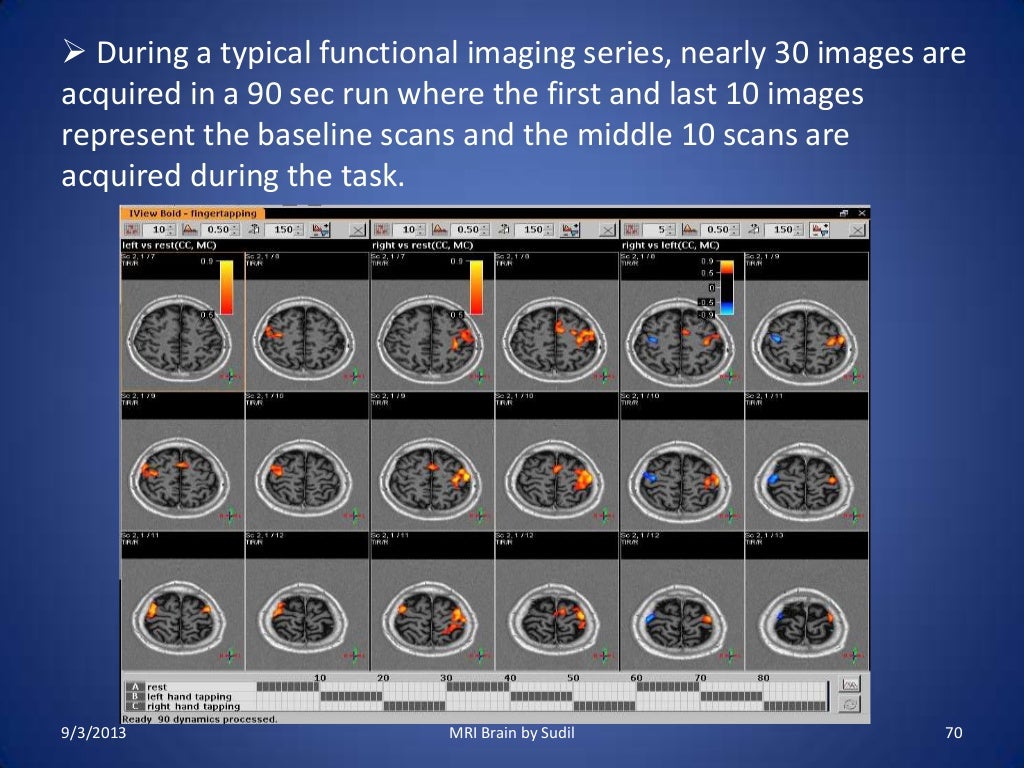
UMD PSYC E-News: RA positions focusing on functional MRI and Autism Spectrum Disorder!
Brain MRI segmentation | Kaggle Journal of Neuro-Oncology, 2017. This dataset contains brain MR images together with manual FLAIR abnormality segmentation masks. The images were obtained from The Cancer Imaging Archive (TCIA). They correspond to 110 patients included in The Cancer Genome Atlas (TCGA) lower-grade glioma collection with at least fluid-attenuated inversion ...
A Hybrid Approach for Automatic Classification of Brain MRI Using Genetic Algorithm and Support ...
Frontiers | 101 Labeled Brain Images and a Consistent Human Cortical ... Labeled anatomical subdivisions of the brain enable one to quantify and report brain imaging data within brain regions, which is routinely done for functional, diffusion, and structural magnetic resonance images (f/d/MRI) and positron emission tomography data.

Mouse brain seen in sharpest detail ever | Kurzweil
matlab - Brain MRI Segmentation Using FCM (Labeling) - Stack Overflow Brain MRI Segmentation Using FCM (Labeling) I am doing Brain MRI segmentation using Fuzzy C-Means, The volume image is n slices, and I apply the FCM for each slice, the output is 4 labels per image (Gray Matter, White Matter, CSF and the background), how I can give the same label (Color) for each material for all the slices) I am using matlab.

Medical Legal Demonstrative Evidence
101 labeled brain images and a consistent human cortical labeling ... given how difficult it is to label brains, the mindboggle-101 dataset is intended to serve as brain atlases for use in labeling other brains, as a normative dataset to establish morphometric variation in a healthy population for comparison against clinical populations, and contribute to the development, training, testing, and evaluation of …

Radiology MRI: Neonatal Intraventricular Hemorrhage
MRI Brain Animated Quiz - University of Minnesota Note: spacebar toggles labels; also arrow keys do Previous/Next Sequentially click/tap: first the dot associated with a term; then, its corresponding target dot on the MRI image. If a line connection appears, your choice was correct!
Brain Anatomy Differences Between Autistic and Typically Developing Individuals Are ...
NITRC: Manually Labeled MRI Brain Scan Database: Tool/Resource Info This is a continuously growing and improving database of high-quality neuroanatomically labeled MRI brain scans, created not by an algorithm, but by neuroanatomical experts. All results are checked and corrected. Regions of interest include the usual sub-cortical structures (thalamus, caudate, putamen, hippocampus, etc), along with ventricles ...

Gamma Knife for Trigeminal Neuralgia
Labeled imaging anatomy cases | Radiology Reference Article ... This article lists a series of labeled imaging anatomy cases by body region and modality. Brain CT head: non-contrast axial CT head: non-contrast coronal CT head: non-contrast sagittal CT head: angiogram axial CT head: angiogram coronal CT...
MRI Procedure of Brain
101 Labeled Brain Images and a Consistent Human Cortical Labeling ... Labeled anatomical subdivisions of the brain enable one to quantify and report brain imaging data within brain regions, which is routinely done for functional, diffusion, and structural magnetic resonance images (f/d/MRI) and positron emission tomography data.

Neuroanatomy - encyclopedia article - Citizendium
CaseStacks.com - MRI Brain Anatomy Labelled radiographs and CT/MRI series teaching anatomy with a level of detail appropriate for medical students and junior residents. Pelvis Pelvic MRI anatomy Chest Chest radiograph & CT anatomy Body Abdominal CT anatomy Cardiac Cardiac CT anatomy Brain Brain & calvarial anatomy on CT/MRI Cranial Nerves Cranial nerves on MRI

MRI Brain Poster | Zazzle.com
Atlas of BRAIN MRI - W-Radiology Brain magnetic resonance imaging (MRI) is a common medical imaging method that allows clinicians to examine the brain's anatomy (1). It uses a magnetic field and radio waves to produce detailed images of the brain and the brainstem to detect various conditions (2).
Gamma Knife for Trigeminal Neuralgia

MRI of Brain - Stock Image - M134/0816 - Science Photo Library

MRI Brain Planning

February | 2011 | Thought Broadcast
Post a Comment for "41 brain mri with labels"