38 knee joint with labels
Label The Structures Of The Knee. Chegg - Solved Match The Knee Joint ... Structure of the knee joint 1. Ty label the structures of the knee joint (superior view by clicking and dragging the labels to the correct location lateral menis eu synovial . Label the structures of the knee. Label the structures of the knee. Tibia patellar surface lateral condyle of femur 16 medial condyle of femur anterior cruciate . Knee Joint Anatomy: Structure, Function & Injuries - Knee Pain Exp The specific design of knee joint anatomy allows a number of functions: Supports the body in upright position without muscles having to work. Helps in lowering and raising body e.g. sitting, climbing and squatting. Allows rotation/twisting of the leg to place and position foot accurately.
shoulder joint with labels - Scottsdale Joint Center The Scottsdale Joint Center is in Arizona - Call us at 480-994-1149. Dr. Stuart Kozinn is an orthopedic surgeon in private practice in Scottsdale.

Knee joint with labels
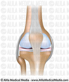
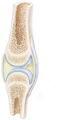
A Diagrammatic Explanation of the Parts of the Human Knee Knee actually consists of three bones - femur, tibia and patella. Femur is the thigh bone, tibia is the shin bone and patella is the small cap like structure which rests on the other two bones. Femur is considered as the largest bone in the human body. The femur and the tibia meets at the tibiofemoral joint and patella rests on top of this joint. Labeling the Knee Joint Quiz - PurposeGames.com This is an online quiz called Labeling the Knee Joint There is a printable worksheet available for download here so you can take the quiz with pen and paper. Your Skills & Rank Total Points 0 Get started! Today's Rank -- 0 Today 's Points One of us! Game Points 11 You need to get 100% to score the 11 points available Actions Alila Medical Media | Knee joint, basic labels | Medical illustration Image size: 39.1 Mpixels (112 MB uncompressed) - 6250x6250 pixels (20.8x20.8 in / 52.9x52.9 cm at 300 ppi)
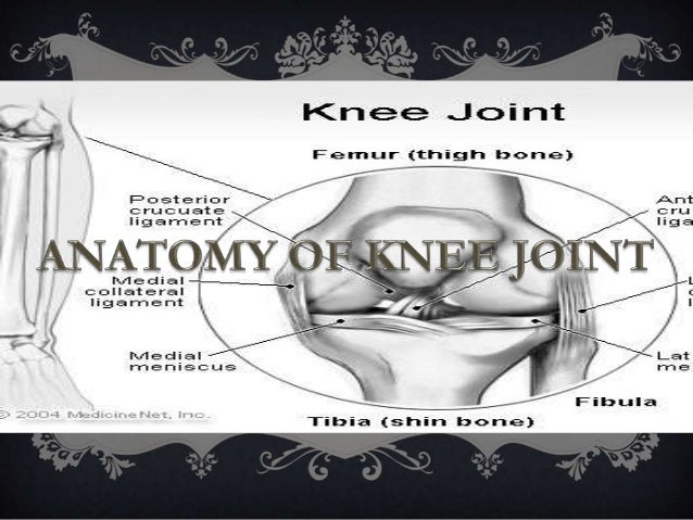
Knee joint with labels. Automatic Atlas-based Three-label Cartilage Segmentation from MR Knee ... Since femoral and tibial cartilage may touch, a joint segmentation using a multi-class segmentation is desirable. We make use of a three-label segmentation approach ensuring distinct labels for touching objects. To customize the method for cartilage segmentation, we incorporate an anisotropic regularization term into the three-label ... Label The Structures Of The Knee. - New Philippines expressways being ... Label the structures of the knee. To deepen the articular surface of the tibia, . The posterior and anterior cruciate ligaments (pcl and acl) limit forward motion of the knee bones, keeping them stable. The 3b scientific® anatomy video knee joint demonstrates the structure of the knee joint. Knee Anatomy: Bones, Muscles, Tendons, and Ligaments Bones Around the Knee There are three important bones that come together at the knee joint: The tibia (shin bone) The femur (thigh bone) The patella (kneecap) A fourth bone, the fibula, is located just next to the tibia and knee joint, and can play an important role in some knee conditions. Knee (Human Anatomy): Function, Parts, Conditions, Treatments - WebMD The knee is one of the largest and most complex joints in the body. The knee joins the thigh bone (femur) to the shin bone (tibia). The smaller bone that runs alongside the tibia (fibula) and the...
label the knee Quiz - PurposeGames.com An unregistered player played the game 2 days ago About this Quiz This is an online quiz called label the knee There is a printable worksheet available for download here so you can take the quiz with pen and paper. Your Skills & Rank Total Points 0 Get started! Today's Rank -- 0 Today 's Points One of us! Points 13 to score the 13 points available Knee joint: anatomy, ligaments and movements | Kenhub Mar 03, 2022 · The tibiofemoral joint Medial condyle of femur Condylus medialis femoris 1/7 The tibiofemoral joint is an articulation between the lateral and medial condyles of the distal end of the femur and the tibial plateaus, both of which are covered by a thick layer of hyaline cartilage . The knee (MRI): Atlas of anatomy in medical imagery - IMAIOS Anatomy of the knee on a coronal slice (MRI) : meniscus (lateral and medial), cruciate ligaments, vastus (lateralis, intermedius, medialis), tibial and fibular collateral ligaments. On "Contrast" the user can choose the type of MRI sequence: spin-echo T1 or proton-density with fat saturation sequences. On "Series" it is possible to ... Knee Joint Picture Image on MedicineNet.com The knee functions to allow movement of the leg and is critical to normal walking. The knee flexes normally to a maximum of 135 degrees and extends to 0 degrees. The bursae, or fluid-filled sacs, serve as gliding surfaces for the tendons to reduce the force of friction as these tendons move. The knee is a weight-bearing joint.
Anatomy of human knee joint with labels — Stock photos "Anatomy of human knee joint with labels" is an authentic stock image by StocktrekImages. It's available in the following resolutions: 1049 x 1600px, 1704 x 2600px, 3422 x 5220px. The minimum price for an image is 49$. Image in the highest quality is 3422 x 5220px, 300 dpi, and costs 449$. Similar Images Same Series Keywords Text Bones Knee Joint - label pictures Flashcards | Quizlet Knee Joint - label pictures STUDY Flashcards Learn Write Spell Test PLAY Match Gravity Created by cfreynolds2018 Terms in this set (7) 1. Femur 2. Articular capsule 3. PCL 4. Lateral Meniscus 5. ACL 6. Tibia 1-6 7. Quadracep tendon 8. Suprapatellar bursa 9. Patella 10. Subcutaneous prepatellar bursa 11. Synovial cavity 12. Lateral Meniscus 13. The Knee Joint - Articulations - Movements - TeachMeAnatomy The knee joint is a hinge type synovial joint, which mainly allows for flexion and extension (and a small degree of medial and lateral rotation). It is formed by articulations between the patella, femur and tibia. In this article, we shall examine the anatomy of the knee joint - its articulating surfaces, ligaments and neurovascular supply. Joint label Images, Stock Photos & Vectors | Shutterstock 25,666 joint label stock photos, vectors, and illustrations are available royalty-free. See joint label stock video clips ... Knee joint anatomy labeled. Types of ...
Alila Medical Media | Knee joint labeled. | Medical illustration
Knee Anatomy, Diagram & Pictures | Body Maps - Healthline The knee is the meeting point of the femur (thigh bone) in the upper leg and the tibia (shinbone) in the lower leg. The fibula (calf bone), the other bone in the lower leg, is connected to the...
34 Label Knee Joint - Labels Information List
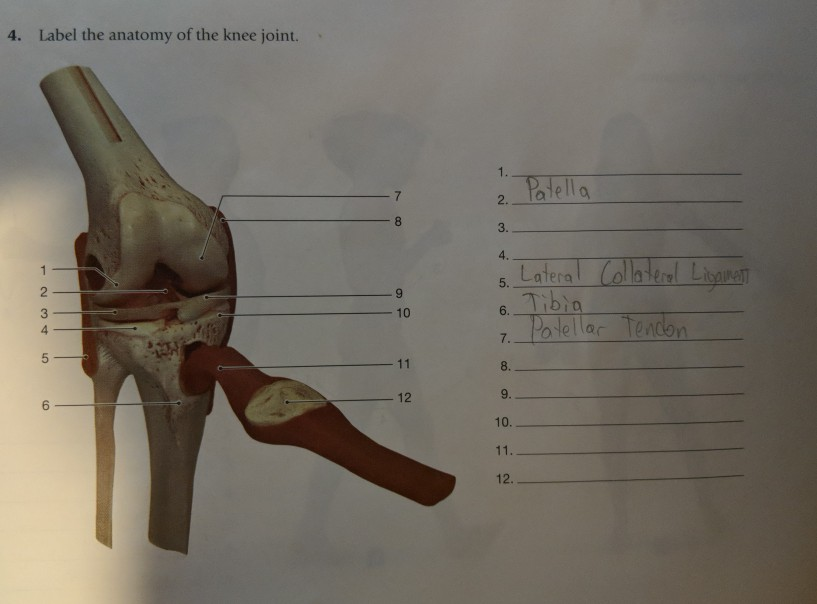
Solved 3 of 5 B. Structure of the knee joint 1. Label the - Chegg Label the parts of the knee joint models anterior cruciate ligament, femur, fibula, fibular collateral ligament, meniscus, patella, patellar ligament, posterior cruciate ligament, tendon of the quadriceps, tibia, tibial collateral ligament 2. Give the functions of the following structures often found in a synovial This problem has been solved!

Health Leads UK Ltd: Supplements for Joint Health
Knee Joint Labeled Diagram stock vector. Illustration of arthritis ... Osteoarthritis of knee joint. Knee-joint side view. Knee joint pain, burning knee. Human knee joint 3d model vector illustration. Low poly design future technology cure pain treatment. Blue background. Joint icon. Medical infographic orthopedic.

Medical suggestions
Knee Joint - San Diego Mesa College Knee Joint. Click on a photo for a larger view of the model. Click on L abel for the labeled model. Back to Muscular System. Anterior: Anterior without patella: Posterior: Label: Label: Label : Label: Label:

Blog not found
Knee Joint Label Flashcards | Quizlet Knee Joint Label STUDY Flashcards Learn Write Spell Test PLAY Match Gravity Created by LaLaKub91 Terms in this set (10) femur What is A? lateral collateral ligament what is d? lateral meniscus what is e? fibula what is g? tibia what is h? posterior cruciate ligament What is j? anterior cruciate ligament what is k? medial meniscus what is l?
Print A&P Chapter 8 Joints flashcards | Easy Notecards
A Labeled Diagram of the Knee With an Insight into Its Working Labeled Diagram of the Knee Joint Knee joint is one of the most important hinge joints of our body. Its complexity and its efficiency is the best example of God's creation. The anatomy of the knee consists of bones, muscles, nerves, cartilages, tendons and ligaments. All these parts combine and work together.

Alila Medical Media | Knee joint labeled drawing. | Medical illustration
Knee x-ray - labeling questions | Radiology Case | Radiopaedia.org Normal X-ray Knee - Frontal (with labels) Annotated image Annotated image Frontal Knee Frontal 1. Femoral shaft 2. Patella 3. Base of patella 4. Apex of patella 5. Adductor tubercle of femur 6. Medial epicondyle of femur 7. Medial condyle of femur 8. Lateral epicondyle of femur 9. Lateral condyle of femur 10. Groove for popliteus 11.

Human Anatomy Lab: Knee Joint Model
Amazon.com: anatomical model knee Axis Scientific Functional Knee Model - Anatomically Correct Knee Joint with Life Like Ligaments That Can Show Movement, Includes Base, Detailed Full Color Product Manual, Worry Free 3 Year Warranty 22 $49 99 Get it as soon as Wed, Apr 13 FREE Shipping by Amazon
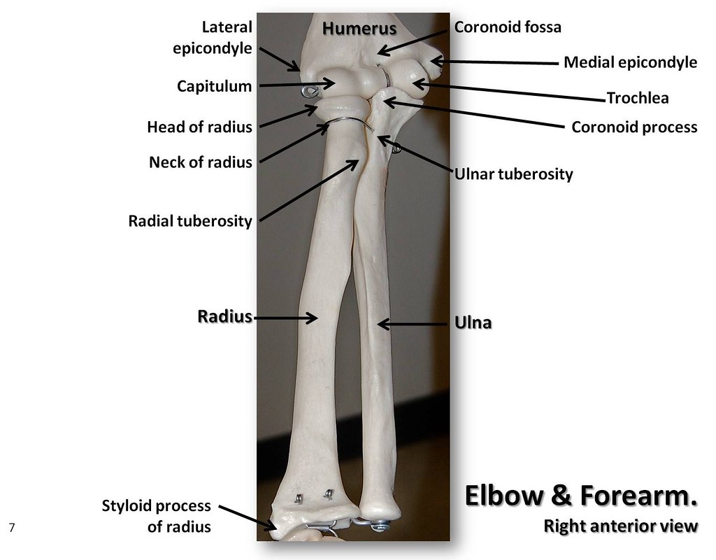
Bones of the elbow and forearm, anterior view with labels … | Flickr
Solved Correctly label the following anatomical features of - Chegg Question: Correctly label the following anatomical features of the knee joint. Patellar ligament Synovial membrane Articular capsule Articular cartilage Fat pad Joint cavity This problem has been solved! See the answer Show transcribed image text Expert Answer 100% (1 rating) Articular capsule. Articular … View the full answer

Total Knee Replacement of the Right Knee | Total knee replacement, Knee replacement, Knee ...
Label The Structures Of The Knee Joint - Solved Procedure 1 Identifying ... The knee joint is essentially made up of three bones: Start studying knee joint label. The femur (thigh bone), tibia (shin bone), and patella (kneecap) make up the bones of the knee. The medial and lateral menisci are fibrocartilage structures in the knee that serve two functions: The knee joint has three parts. The knee joint keeps these bones ...

Osteoarthritis of knee joint Poster | Zazzle.com
Knee Joint - Anatomy Pictures and Information - Innerbody Jul 16, 2019 · The knee, also known as the tibiofemoral joint, is a synovial hinge joint formed between three bones: the femur, tibia, and patella. Two rounded, convex processes (known as condyles) on the distal end of the femur meet two rounded, concave condyles at the proximal end of the tibia. Continue Scrolling To Read More Below... Additional Resources
KNEE JOINT
Alila Medical Media | Knee joint, basic labels | Medical illustration Image size: 39.1 Mpixels (112 MB uncompressed) - 6250x6250 pixels (20.8x20.8 in / 52.9x52.9 cm at 300 ppi)

Alila Medical Media | Knee joint, basic labels | Medical illustration
Labeling the Knee Joint Quiz - PurposeGames.com This is an online quiz called Labeling the Knee Joint There is a printable worksheet available for download here so you can take the quiz with pen and paper. Your Skills & Rank Total Points 0 Get started! Today's Rank -- 0 Today 's Points One of us! Game Points 11 You need to get 100% to score the 11 points available Actions
Knee joint
A Diagrammatic Explanation of the Parts of the Human Knee Knee actually consists of three bones - femur, tibia and patella. Femur is the thigh bone, tibia is the shin bone and patella is the small cap like structure which rests on the other two bones. Femur is considered as the largest bone in the human body. The femur and the tibia meets at the tibiofemoral joint and patella rests on top of this joint.

Human Anatomy Lab: Knee Joint Model
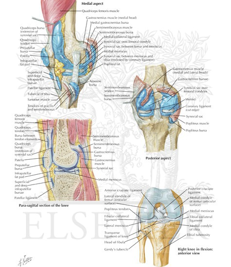
Welcome To Netter Images
Post a Comment for "38 knee joint with labels"